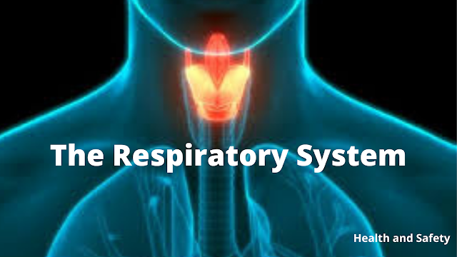
The Respiratory System of Human Body
We are going to discuss about the respiratory system in this article. One of the most vital organ systems of our body is the respiratory system. We breathe about 16,000 to 24,000 times per day, which exchanges about 11,000 liters of air with the atmosphere in this process. The oxygen of the atmospheric air is delivered to the human body, which is then utilized by all the tissues, and the carbon dioxide which is produced by the human body is exported to the lungs and then exchanged with the atmospheric air,
In this article we will first talk about the brief anatomy and physiology of the human respiratory system and then we will discuss how the gas transport to the alveoli occurs, we will talk about how the human respiratory system works together with the circulatory system and then we will talk about how the gas exchange happens in the alveoli.
So, first coming to the anatomy and physiology of the human respiratory system, the vocal cords present in the larynx divide the respiratory tract into the upper respiratory tract and the lower respiratory tract. The upper respiratory tract contains structures like the nasal cavity, the pharynx and the part of the larynx above the vocal cords, the lower respiratory tract starts with the part of the larynx below the vocal cords, trachea, bronchi, bronchioles as well as the alveoli.
So let's first talk about the brief anatomy of the nose and the nasal cavity, now if we take a look at this diagram in a bit of detail we can see that the nasal cavity is made up of a roof, which is in turn made up of the bones forming the base of the skull, it also consists of a floor which is made up of the palatine bones. The main job of the nose and the nasal cavity in the respiratory tract is the filtration of air, this is made possible by small minute hair-like structures present in the nasal cavity called cilia. The filtration of air is also facilitated by the mucus which is secreted by the nasal walls; this traps dust pulls in as well as other pollutants present in the atmospheric air. The nasal cavity warms and humidifies the air, which prevents the dryness in the respiratory membranes.
Oral cavity also serves as a secondary opening for the respiratory tract, the downside of this is that there is no filtration no humidification, and no temperature regulation through the oral cavity. The plus point is that the oral cavity has a wider opening so it facilitates the greater intake of air during exercise.
So till now, we have discussed the air that we inhale passes through our nasal cavity or the oral cavity into the pharynx, so coming to the next important structure in the human respiratory tract which is the pharynx.
Pharynx is basically a muscular tube which lies behind the nose in the mouth as well as the larynx and it connects the oral and the nasal cavity to the larynx as well as the esophagus. The part you see is the part which is called the nasopharynx this is the part which lies behind the nasal cavity. The second part of the pharynx is called the oropharynx, which is the part lies behind the oral cavity, and the third is called the laryngopharynx which lies behind the larynx.
So the air that we inhale through our nose or to our oral cavity passes back into the pharynx and when the air reaches the pharynx it has two important structures where it can go forward. The first one is the trachea which lies in the front and second one is the esophagus which lies to the back.
Normally the air enters in the trachea because esophagus at normal conditions is a collapsed structure. One important structure to note here is the epiglottis, the epiglottis is basically a cartilaginous structure which closes the laryngeal inlet and prevents entry of food into the trachea and urther the lungs.
The next important structure in the respiratory tract is the larynx. The larynx is located right below the pharynx and it is also known as the voice box as well as the Adam's apple the larynx is made up of many cartilages and the larynx contains the vocal cords the main functions of the larynx in the human respiratory system is that it connects the pharynx to the trachea and the second important structure is the production of sound or speech.
The next important structure is the trachea. So the trachea is basically a tube-like structure which connects the larynx to the bronchi and the bronchi in turn connect to the lungs. If we take a closer look at the section of the trachea we can see that the trachea is basically made up of C-shaped cartilages from above to below. These cartilages basically prevent the collapse of the trachea because there is a negative pressure in trachea as well as the lungs during inhalation. If you see closely these tracheal rings are basically C-shaped, these are not completely circular because esophagus lies behind the trachea and if these rings were circular they would have compressed to the esophagus during swallowing, and this could have led to choking.
The trachea then divides into the primary bronchi, now this is called the primary bronchi because it is the first division of the trachea, the primary bronchi then divided into secondary bronchioles and then the secondary bronchioles divide into the tertiary bronchioles. What happens next is that these tertiary bronchioles again divided for about 20-23 divisions to form the conducting bronchioles. The tertiary bronchioles divided many times to form very small sac-like structures, the tertiary bronchioles lead into the conducting bronchioles, the conducting bronchioles then form a 4 to 5 division. These respiratory bronchioles are in turn connected to the alveoli.
The alveoli are sac-like structures in which the actual gas exchange happens. The conducting bronchioles are called conducting bronchioles because no gas exchange happens in them. Since they have a large or thick walls but respiratory bronchioles have very thin walls and some amount of gas exchange can happen in these respiratory bronchioles, but majority of the gas exchange happens in the alveoli because of their close proximity to the blood vessels.
The division of the bronchioles to such an extent increases the effective surface area of the lungs so the air that we inhale finally reaches into the alveoli, The lungs, the heart as well as a circulation pool of the body, showing the deoxygenated blood in blue color as well as the oxygenated blood in the red color. The heart receives all the deoxygenated blood from the body through the superior as well as the inferior vena cava, this blood has a low concentration of oxygen and this is then pumped to the lungs through the pulmonary arteries, in the lungs this deoxygenated blood is subjected to gas exchange which converts it into oxygenated blood which is pumped back to the heart.The heart then pumps this oxygenated blood to the body which utilizes the oxygen present in the blood and again this blood is converted into deoxygenated blood which is pumped back to the heart. The heart pumps all the deoxygenated blood to the lungs through two main arteries the right and the left pulmonary artery, just as we saw with the previous branching pattern that a single bronchus almost divides 20 to 25 times before the formation of alveoli. The similar phenomena happens with the large blood vessels that enter into the lungs, these blood vessels also divide several times which leads to formation of small capillaries that are in extremely close contact with the alveoli.
Due to this the cardiac output that comes in the large vessels divides into many small streams of blood. This leads to exposure of the 5 liters of blood coming through the heart to almost 250 to 300 million alveoli per minute. Due to this a rapid gaseous exchange happens between the alveolar air and the blood that is exposed to the alveoli.
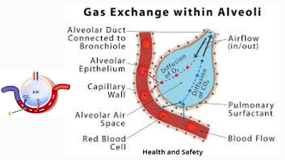
Now let's try to understand how the actual gas transport happens inside single alveoli, the air that we inhale passes through a respiratory tract deep into our body into a bunch of structures called alveoli, well this is important to create a very less distance between the alveolar air and the blood.
To understand this take a closer look at this section of an alveoli which are attached to the end of a respiratory bronchioles. You can see that a bunch of alveoli are surrounded closely by blood vessels. If we take a single alveoli out of this bunch and magnify it under microscope we can see that the alveoli is completely surrounded by a single capillary but in reality they are surrounded by multiple capillaries to have very close contact with the blood.
In these capillaries the deoxygenated blood enters from one side it exchanges the gases with the alveolar air and finally oxygenated blood is released from the other side. If we take a closer look at the junction between the alveoli and the capillary wall. On the other side we have the blood vessels which contain the RBC's, and the wall of the blood vessel is the endothelium and between them is the basement membrane which separates these two cell layers.
So you can see clearly that the air that we inhale in the alveoli is a very close proximity which is approximately few micrometers from the blood vessel. Here comes a roll of difference in the concentration of gases on either side most of you know that the diffusion occurs due to the difference in concentration of a substance on either side.
So if we take a look at the concentration difference of the alveolar air and the blood of carbon dioxide and oxygen, we can see that the alveolar air has a low concentration of carbon dioxide as compared to the blood which has a higher concentration. Whereas opposite of that is true in case of oxygen where high concentration of oxygen is present in alveolar air as compared to low concentration of oxygen in the blood.
Due to this the high concentration of carbon dioxide is exchanged with the alveolar air and the carbon dioxide is released outside in the atmosphere. Whereas the fresh oxygen that is coming through the alveolar air is transported to the blood and blood is made oxygenated.
So the blood coming on one side of the capillary which is a deoxygenated blood is converted into oxygenated blood. On the other side this oxygenated blood is then sent to the heart and the heart then pumps this oxygenated blood to whole of the body which is then utilized. So this was a brief description of the human respiratory system.


.png)

.png)

Comments
Post a Comment